Received: Tue 01, Jul 2025
Accepted: Fri 25, Jul 2025
Abstract
Background: Spinal cord injury (SCI), a profoundly disabling disorder of the central nervous system, involves programmed cell death as a fundamental mechanism underlying neurological deterioration. This study establishes a comprehensive knowledge framework through bibliometric analysis and scientific visualization of existing literature.
Methods: Based on WoSCC, Scopus and PubMed databases to search related literature in the last 10 years, 2403 target documents were screened for bibliometric and visualization analysis using Citespace 6.3.R1 and Bibliometrix.
Results: The analysis have revealed a sustained upward trajectory in annual publication volume within this domain, juxtaposed against a marked decline in citation frequency post-2019. A scholarly workforce comprising 9,333 authors demonstrated limited global interconnectivity, evidenced by an international collaboration index (10.99%) predominantly confined to institutional partnerships. Molecular Neurobiology emerged as the predominant contributor by publication volume, whereas the Journal of Neurotrauma demonstrated both the highest localized citation impact (LCS) and field-specific influence metrics. Dr. Jian Xiao was identified as the most prolific contributor, with Wenzhou Medical University and Jinzhou Medical University serving as foremost institutional contributors. China manifested as the undisputed dominant force across all evaluative dimensions.
Lexical analysis identified three cardinal keyword clusters: apoptosis, spinal cord injury, and activation. Thematic deconstruction via clustering algorithms delineated five principal research domains:
i) Hereditary spastic paraplegia pathogenesis. ii) Mesenchymal stromal cell-mediated regenerative paradigms (Stem cell therapies). iii) NLRP3 inflammasome-oxidative stress axis modulation (Inflammation control). iv) Immunoregulatory pathway engineering (Anti-inflammatory pathways). v) Axonogenesis mechanistic frameworks (Nerve fiber regeneration).
Emerging research frontiers center on:
i) Autophagy flux modulation (augmenting autophagy). ii) Nanotherapeutic delivery systems (hydrogel matrices, polymeric nanoparticles). iii) Ferroptosis inhibition strategies (deferoxamine-mediated iron chelation).
Conclusion: This investigation systematically delineates the knowledge architecture of programmed cell death associated spinal cord injury research, revealing China's quantitative leadership while highlighting critical deficiencies in transnational collaborative frameworks and high-impact scholarly output. Future investigations must prioritize: i) precision targeting of neuroinflammatory cascades (Targeted anti-inflammatory therapies), ii) innovation in bioengineered therapeutic delivery platforms (Advanced drug delivery systems), and iii) clinical translation pipelines, concurrently fostering cross-disciplinary consortia to address diminishing citation impact metrics. Methodological constraints stem from monodirectional data sourcing and inherent temporal latency in bibliometric capture (Clinical testing).
Keywords
Spinal cord injury, programmed death, knowledge graph, research trends, bibliometrics
1. Introduction
Spinal Cord Injury (SCI) is a serious neurological condition in which the structure or function of the spinal cord is impaired due to trauma or disease, resulting in sensory, motor or autonomic dysfunction [1]. In spinal cord injury, programmed cell death is an important mechanism leading to neurologic dysfunction and exacerbation of the injury, for example: Apoptosis: Mitochondrial-driven cell dismantling; Pyroptosis: Inflammasome-induced cell rupture; Ferroptosis: Iron-dependent lipid peroxidation; Cuproptosis: Copper-triggered protein aggregation. Apoptosis exacerbates neuronal and glial cell death during secondary damage after injury, whereas pyroptosis exacerbates the neuroinflammatory response through inflammatory factor release [2]. In addition, iron death is a non-apoptotic cell death caused by iron-dependent lipid peroxidation, and studies have shown that targeted inhibition of iron death can help improve neurological recovery after SCI [3]. In recent years, copper death, a newly discovered mode of programmed death, has also been recognized as possibly playing a role in the pathological process of SCI [4]. An in-depth study of the mechanism of programmed death is important for the development of new neuroprotective strategies.
Bibliometrics is a method that reveals research trends and academic influence in a specific field by quantitatively analyzing indicators such as the number of published scientific literature, citation frequency, and research hotspots. The method can be used to assess the current status of research, identify research frontiers, and provide guidance for future research [5]. Currently, bibliometric studies on SCI are mostly focused on stem cell therapy and other aspects, while systematic metrological analyses on programmed death are still lacking. Bibliometric studies in this field can not only reveal the hotspots and cutting-edge directions of research, but also assess the academic influence of different institutions, authors and journals, and promote scientific cooperation and resource sharing. In addition, this type of research can provide data support for research administrators and funding agencies to help formulate rational research funding policies and development strategies. The visualization tools used in this research are CiteSpace and Bibliometrix in R language.
CiteSpace is a Java-based tool for bibliometric analysis and scientific knowledge mapping developed by Prof. It is mainly used to analyze research trends, hot topics, collaboration networks and knowledge structure evolution in academic literature [6]. CiteSpace helps researchers identify important literature, core authors and their collaborations in the field, and track the evolution of knowledge through methods such as co-citation analysis, keyword co-occurrence analysis, and emergent word detection. The tool is particularly suitable for analyzing dynamic changes in scientific fields, such as the identification of research frontiers, visualization of hotspots in the field, and the construction of scientific collaboration networks. In addition, CiteSpace provides a variety of visualization features, such as timeline views, cluster diagrams, and centrality analyses, to help researchers more intuitively understand the structure and trends of literature data.
Bibliometrix is a public source R-based bibliometric analysis package developed by Prof. Massimo Aria and his team at the University of Naples Paternope, Italy. Bibliometrix provides a complete set of features for bibliometric analysis, including bibliometric data cleaning, descriptive statistics, co-word analysis, co-citation analysis, scientific collaboration analysis, research topic clustering, and knowledge structure visualization. The companion Biblioshiny is a user-friendly web interface based on the Shiny framework that enables researchers to perform complex bibliometric analyses without writing R code. Bibliometrix supports data import from a variety of academic databases (e.g., Web of Science, Scopus, and PubMed) and provides powerful visualization features such as three-field mapping, topic evolution analysis, and literature coupling analysis. Due to its flexibility and extensibility, Bibliometrix is widely used in bibliometric studies, especially for researchers who need to customize the analysis process and in-depth statistical analysis [7].
2. Methods
2.1. Data Retrieval and Collection
After determining the purpose of the study, we identified the keywords needed for the search: (“programmed cell death” OR apoptosis OR parthanatos OR necroptosis OR pyroptosis OR autophagy OR “cell death pathway” OR “regulated cell death” OR ferroptosis) AND (”spinal cord injury” OR ‘spinal cord trauma’ OR paraplegi* OR tetraplegi*). After discussion, we finally decided to use Web of Science Core Collection (WoSCC), Scopus and PubMed as source databases for the search.
The time interval of 2015-01-01 to 2025-01-01 was used as the premise, and the corresponding search formula was entered in each database:
Enter the identified search formula TS= ((“programmed cell death” OR apoptosis OR parthanatos OR necroptosis OR pyroptosis OR autophagy OR “cell death pathway” OR “regulated cell death” OR ferroptosis) AND (” spinal cord injury” OR ‘spinal cord trauma’ OR paraplegi* OR tetraplegi*)), yielding 2914 results.
In Scopus, enter the search formula TITLE-ABS- KEY((“programmed cell death” OR apoptosis OR parthanatos OR necroptosis OR pyroptosis OR autophagy OR “cell death pathway” OR “regulated cell death” OR ferroptosis) AND (“spinal cord injury” OR 'spinal cord trauma' OR paraplegi* OR tetraplegi*)), yielding 335 results.
Entering the search formula in PubMed (“programmed cell death” [Title/Abstract] OR apoptosis [Title/Abstract] OR parthanatos [Title/Abstract] OR necroptosis [Title/ Abstract] OR pyroptosis [Title/Abstract] OR autophagy [Title/Abstract] OR “cell death pathway” [Title/Abstract] OR “regulated cell death”[Title/Abstract] OR ferroptosis[Title/Abstract]) AND (“spinal cord injury”[Title/Abstract] OR 'spinal cord trauma'[Title/Abstract] OR paraplegi*[Title/Abstract] OR tetraplegi*[Title/Abstract]), yielding 2111 results.
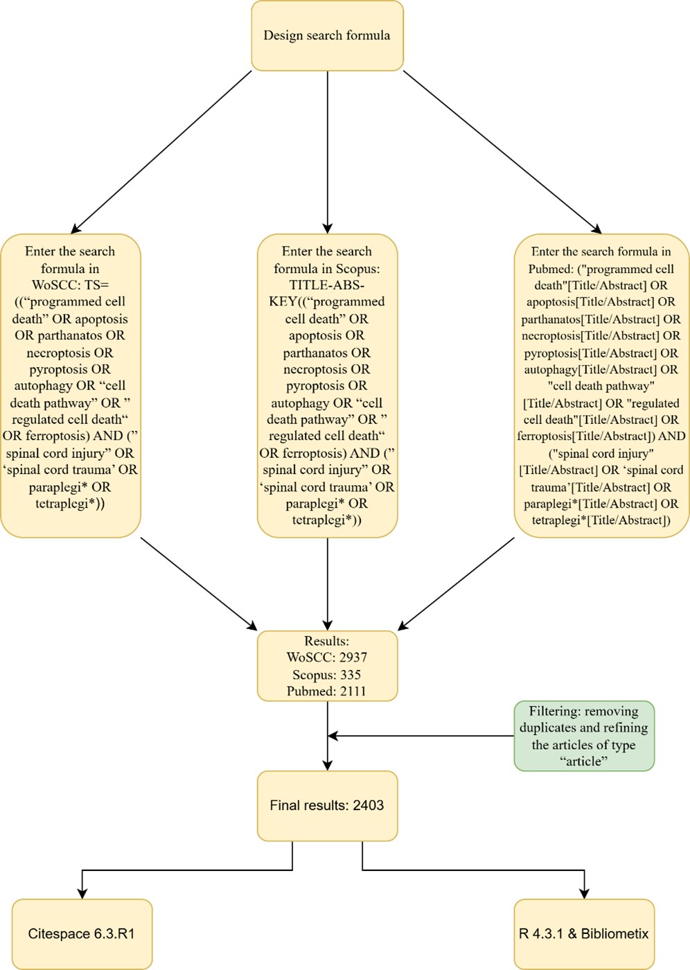
Each of the three databases was searched by three authors and screened according to the same principles. In this study, our team agreed that except for the literature of type “article” which is of practical reference significance, the inclusion of other types of literature may cause a large error, so only articles of type “article” were included. Removal of duplicate literature was then carried out independently by two authors. The results of the two authors were compared and discussed to decide the final literature to be included in the study. It is necessary to state that no information was obtained during or after the retrieval that could identify the individual participant. The general process is shown in (Figure 1).
2.2. Data Analysis
The collected literature was analyzed using citespace 6.3.R1, and the plain text file exported from WoSCC was first de-duplicated, a step that prevents duplicate entries due to operational errors or other reasons during data export. The result of de-duplication showed no duplicates. Create a new project, select "From 2015, JAN to 2025, JAN" for the time interval, select 1 for Years Per Slice, and leave the rest as default, and select any item in Node Types for visual analysis. Bibliometrix was loaded with R 4.3.1 to analyze the literature, and it can be downloaded from the official website (Link), firstly, the plain text file was compressed into a Zip file and imported into Bibliometrix, keeping the default settings, and running the program under each project to get the corresponding results. Detailed operations are available in the respective manuals.
3. Results
3.1. Overview of the Field
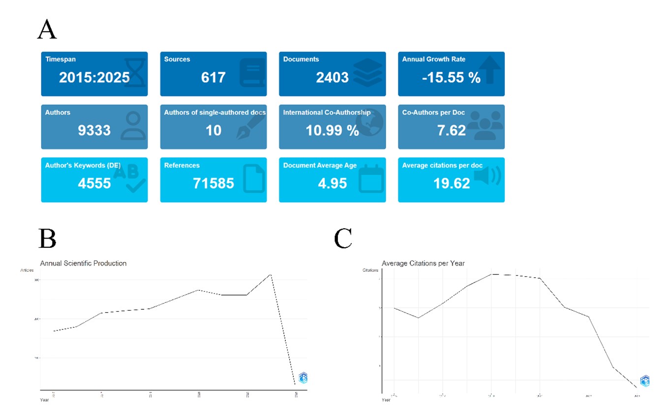
The time span is 2015-2025, with a total of 2403 publications from 617 journals, and the annual growth rate of -15.55% is due to the fact that the time cutoff is 2025-01-01, and the literature from 2025 was not included to cause this result. A total of 9333 authors were involved in the study, with an average of 7.62 authors per document and only 10 single author documents, indicating that research into this field is highly dependent on teamwork. The rate of international collaboration between authors was 10.99%, indicating that most of the studies failed to get help internationally. There were 4555 keywords for authors, indicating the diversity of the field. The average age of the literature is 4.95 years, reflecting the high activity of the field in the last five years, and the average number of citations per article is higher than most biomedical fields at 19.62, suggesting high quality research or the presence of landmark literature (Figure 2A). The annual scientific output shows a steady increase in the literature from 2015-2021, a small decrease in the literature after 2021-2023, and a new record in the annual number of publications in 2024 (Figure 2B). The annual average citations in 2015 was 3, and after a decline in 2016 continued to rise until 2019, when it reached its highest, followed by a yearly decline (Figure 2C). We hypothesize that there are three possibilities for this phenomenon: i) most of the subsequent studies lacked innovation resulting in insufficient attention; ii) a shift in research direction resulted in fewer citations to previous articles; and iii) newly published literature is still in the window period and has not yet accumulated enough citations.
3.2. Resources
Leading the way from a publication volume perspective are Molecular Neurobiology (n=80), Neural Regeneration Research (n=73), Molecular Medicine Reports (n=48). The top ten journals in terms of publications are shown in (Figure 3A). It is clear to see that molecular neurobiology and neural regeneration research break ahead of other journals. In the ranking of high locally cited journals J Neurotraum (Journal of Neurotrauma) was ranked first (n=3369), followed by J Neurosci (Journal of Neuroscience) (n=2461), EXP NEUROL (Experimental Neurology) (n=2060) and others (Figure 3B). Of these, only Journal of Neurotrauma appears in both lists, indicating that this journal is not only happy to receive articles in this field, but also demands high quality. Journal of Neurotrauma is unsurprisingly ranked first in terms of local impact of journals according to the H-index. Figure 3C shows the top ten journals in the H-index, and the top three are Molecular Neurobiology, Neural Regeneration Research in addition to Journal of Neurotrauma. Table 1 shows more detailed information about them, including H index, G index, M index, etc. Figure 3D identifies the core journals in this field, and they are listed in order of the number of publications, such as Molecular Neurobiology, which is ranked first in terms of the number of publications, etc. The list is shown in the (Table 2).
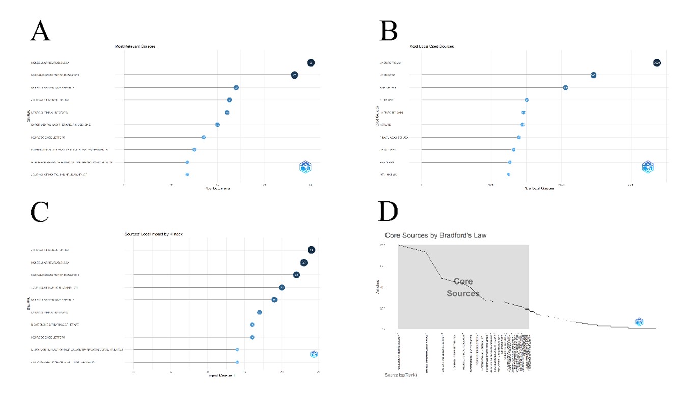
Table. 1. The top ten journals in the H-index and their
G-index, M-index, total citations (TC), number of publications (NP),
publication year start (PY start).
|
Element |
h_index |
g_index |
m_index |
TC |
NP |
PY_start |
|
JOURNAL OF NEUROTRAUMA |
24 |
41 |
2.182 |
1702 |
45 |
2015 |
|
MOLECULAR NEUROBIOLOGY |
23 |
37 |
2.091 |
1575 |
80 |
2015 |
|
NEURAL REGENERATION RESEARCH |
22 |
36 |
2 |
1610 |
73 |
2015 |
|
JOURNAL OF NEUROINFLAMMATION |
20 |
24 |
2 |
1591 |
24 |
2016 |
|
MOLECULAR MEDICINE REPORTS |
19 |
26 |
1.727 |
873 |
48 |
2015 |
|
NEUROCHEMICAL RESEARCH |
17 |
29 |
1.545 |
918 |
44 |
2015 |
|
BIOMEDICINE & PHARMACOTHERAPY |
16 |
24 |
1.6 |
667 |
24 |
2016 |
|
NEUROSCIENCE LETTERS |
16 |
25 |
1.455 |
681 |
34 |
2015 |
|
EUROPEAN REVIEW FOR MEDICAL AND
PHARMACOLOGICAL SCIENCES |
14 |
18 |
1.556 |
420 |
27 |
2017 |
|
OXIDATIVE MEDICINE AND CELLULAR LONGEVITY |
14 |
19 |
1.4 |
540 |
19 |
2016 |
Table. 2. Core journals and related information.
|
SO |
Rank |
Freq |
cumFreq |
Zone |
|
MOLECULAR NEUROBIOLOGY |
1 |
80 |
80 |
Zone 1 |
|
NEURAL REGENERATION RESEARCH |
2 |
73 |
153 |
Zone 1 |
|
MOLECULAR MEDICINE REPORTS |
3 |
48 |
201 |
Zone 1 |
|
JOURNAL OF NEUROTRAUMA |
4 |
45 |
246 |
Zone 1 |
|
NEUROCHEMICAL RESEARCH |
5 |
44 |
290 |
Zone 1 |
|
EXPERIMENTAL AND THERAPEUTIC MEDICINE |
6 |
40 |
330 |
Zone 1 |
|
NEUROSCIENCE LETTERS |
7 |
34 |
364 |
Zone 1 |
|
INTERNATIONAL JOURNAL OF CLINICAL AND
EXPERIMENTAL MEDICINE |
8 |
30 |
394 |
Zone 1 |
|
EUROPEAN REVIEW FOR MEDICAL AND
PHARMACOLOGICAL SCIENCES |
9 |
27 |
421 |
Zone 1 |
|
JOURNAL OF MOLECULAR NEUROSCIENCE |
10 |
27 |
448 |
Zone 1 |
|
CNS NEUROSCIENCE & THERAPEUTICS |
11 |
26 |
474 |
Zone 1 |
|
FRONTIERS IN PHARMACOLOGY |
12 |
26 |
500 |
Zone 1 |
|
NEUROSCIENCE |
13 |
26 |
526 |
Zone 1 |
|
SCIENTIFIC REPORTS |
14 |
26 |
552 |
Zone 1 |
|
INTERNATIONAL JOURNAL OF CLINICAL AND
EXPERIMENTAL PATHOLOGY |
15 |
25 |
577 |
Zone 1 |
|
BIOMEDICINE & PHARMACOTHERAPY |
16 |
24 |
601 |
Zone 1 |
|
JOURNAL OF NEUROINFLAMMATION |
17 |
24 |
625 |
Zone 1 |
|
EXPERIMENTAL NEUROLOGY |
18 |
23 |
648 |
Zone 1 |
|
INTERNATIONAL IMMUNOPHARMACOLOGY |
19 |
22 |
670 |
Zone 1 |
|
INTERNATIONAL JOURNAL OF MOLECULAR SCIENCES |
20 |
22 |
692 |
Zone 1 |
|
BIOCHEMICAL AND BIOPHYSICAL RESEARCH
COMMUNICATIONS |
21 |
21 |
713 |
Zone 1 |
|
JOURNAL OF CELLULAR AND MOLECULAR MEDICINE |
22 |
21 |
734 |
Zone 1 |
|
BRAIN RESEARCH |
23 |
20 |
754 |
Zone 1 |
|
BRAIN RESEARCH BULLETIN |
24 |
19 |
773 |
Zone 1 |
|
JOURNAL OF SPINAL CORD MEDICINE |
25 |
19 |
792 |
Zone 1 |
|
OXIDATIVE MEDICINE AND CELLULAR LONGEVITY |
26 |
19 |
811 |
Zone 1 |
3.3. Authors
According to Bibliometrix, we obtained the relevant information of authors under this field. The most relevant authors with the highest number of publications are MEI XF (Mei, Xifen) and XIAO J (Xiao, Jian. Wenzhou Medical University) (n=59). Mei, Xifen is an author record generated by the WoS algorithm because the author did not register his/her identity in WoS and claimed his/her article, and there are several authors with the same name who have published in this field. In fact, except for Xiao, Jian, whose identity has been confirmed, none of the other 9 authors on the list have been identified (Figure 4A). Similarly, in (Figure 4B), only Xiao, Jian's identity is confirmed. This shows that there are very few authors with high output in this field. The distribution of authors under this field can be visualized in (Figure 4C): the curve drops sharply between 1-2 publications, indicating that the vast majority of authors have only 1-2 publications, and detailed data can be seen in (Table 3). Where 69.4% of authors have 1 publication, and authors with fewer than 3 publications 83.7%, 97.9% with less than 10 publications, and the percentage of authors with more than 24 publications tends to zero. We again analyzed the collaborative network of the authors using CiteSpace (Figure 4D), and as can be seen from the figure, these authors are divided into multiple clearly delimited groups, with strong collaborative relationships within the groups, while the groups are connected through one or two authors.

Table. 3. The number of authors corresponding to
different publication numbers and the percentage of them.
|
Documents written |
N. of Authors |
Proportion of Authors |
|
1 |
6477 |
0.694 |
|
2 |
1338 |
0.143 |
|
3 |
553 |
0.059 |
|
4 |
295 |
0.032 |
|
5 |
192 |
0.021 |
|
6 |
116 |
0.012 |
|
7 |
60 |
0.006 |
|
8 |
62 |
0.007 |
|
9 |
47 |
0.005 |
|
10 |
24 |
0.003 |
|
11 |
24 |
0.003 |
|
12 |
24 |
0.003 |
|
13 |
16 |
0.002 |
|
14 |
16 |
0.002 |
|
15 |
9 |
0.001 |
|
16 |
8 |
0.001 |
|
17 |
11 |
0.001 |
|
18 |
6 |
0.001 |
|
19 |
6 |
0.001 |
|
20 |
7 |
0.001 |
|
21 |
2 |
0 |
|
22 |
5 |
0.001 |
|
23 |
4 |
0 |
|
24 |
8 |
0.001 |
|
25 |
2 |
0 |
|
26 |
2 |
0 |
|
27 |
2 |
0 |
|
28 |
3 |
0 |
|
30 |
1 |
0 |
|
35 |
1 |
0 |
|
38 |
3 |
0 |
|
39 |
1 |
0 |
|
40 |
1 |
0 |
|
46 |
2 |
0 |
|
48 |
1 |
0 |
|
50 |
1 |
0 |
|
53 |
1 |
0 |
|
59 |
2 |
0 |
3.4. Institutions
network, which can be seen in (Figure 5A), where these institutions have close cooperation. The larger the node in the graph, the more often it cooperates with other institutions, such as Wenzhou Medical University (n=125). Other than that, the top ten are Jinzhou Medical University (n=93), Zhejiang University (n=90), Nanjing Medical University (n=71), Nantong University (n=71), Shanghai Jiao Tong University (n=69), Shandong University (n=61), Xi`an University (n=57), Soochow University-China (n=57), Tianjin Medical University (n=56), (n=57), Soochow University-China (n=57), Tianjin Medical University (n=56). The ten universities are all from China, with the most central being Wenzhou Medical University, which is more adept at collaborating with other institutions. Table 4 shows the outbreaks of institutions that existed in the selected intervals that had a sudden increase in the number of citations at a given time, suggesting that a significant article had been published during that time. We are concerned that the strong citation phenomenon continues until 2025 for seven institutions, namely Nanchang University, Southeast University-China, Anhui Medical University, University of Science & Technology of China, Jinan University, Tsinghua University, and Huzhou University, which may have published important results in the recent years. In (Figure 5B), these institutions are categorized by keywords and then presented in the form of a timeline, which shows that most of the institutions are active during 2015-2020, and some of them are still active in recent years, such as Qingdao Municipal Hospital, University of Health & Rehabilitation Sciences, etc. The 8 clusters labeled by these institutions are: i) neurodegeneration, ii) tfeb, iii) er stress, iv) neuroprotective, v) dimerization, vi) flica, vii) vascular injure, viii) lercanidipine.
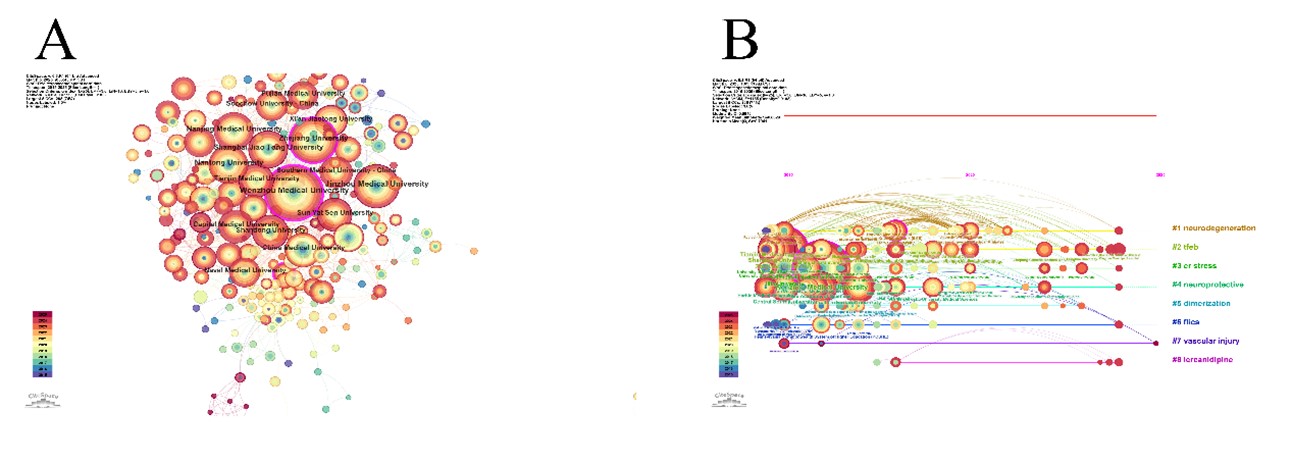
Table. 4. 23 institutions of the citation burst
phenomenon.
|
Top 23 Institutions with the Strongest Citation Bursts |
|||||
|
Institutions |
Year |
Strength |
Begin |
End |
2015 - 2025 |
|
Jilin University |
2015 |
4.7 |
2015 |
2019 |
▃▃▃▃▃▂▂▂▂▂▂ |
|
China Medical University |
2015 |
3.39 |
2015 |
2018 |
▃▃▃▃▂▂▂▂▂▂▂ |
|
Chinese People's Liberation Army General
Hospital |
2015 |
2.9 |
2015 |
2016 |
▃▃▂▂▂▂▂▂▂▂▂ |
|
Kyung Hee University |
2015 |
2.9 |
2015 |
2017 |
▃▃▃▂▂▂▂▂▂▂▂ |
|
Nantong University |
2015 |
2.86 |
2015 |
2016 |
▃▃▂▂▂▂▂▂▂▂▂ |
|
Medical University of South Carolina |
2015 |
2.84 |
2015 |
2016 |
▃▃▂▂▂▂▂▂▂▂▂ |
|
University of Louisville |
2016 |
3.66 |
2016 |
2018 |
▂▃▃▃▂▂▂▂▂▂▂ |
|
Shanghai Jiao Tong University |
2015 |
3.27 |
2016 |
2017 |
▂▃▃▂▂▂▂▂▂▂▂ |
|
Pennsylvania Commonwealth System of Higher
Education (PCSHE) |
2016 |
2.93 |
2016 |
2018 |
▂▃▃▃▂▂▂▂▂▂▂ |
|
Wenzhou University |
2017 |
3.96 |
2017 |
2020 |
▂▂▃▃▃▃▂▂▂▂▂ |
|
Egyptian Knowledge Bank (EKB) |
2018 |
3.13 |
2018 |
2021 |
▂▂▂▃▃▃▃▂▂▂▂ |
|
Southwest Medical University |
2019 |
3.45 |
2019 |
2021 |
▂▂▂▂▃▃▃▂▂▂▂ |
|
Zhengzhou University |
2016 |
3.02 |
2020 |
2021 |
▂▂▂▂▂▃▃▂▂▂▂ |
|
Boston Children's Hospital |
2020 |
2.84 |
2020 |
2021 |
▂▂▂▂▂▃▃▂▂▂▂ |
|
Nanchang University |
2020 |
3.16 |
2021 |
2025 |
▂▂▂▂▂▂▃▃▃▃▃ |
|
Southeast University - China |
2022 |
4.73 |
2022 |
2025 |
▂▂▂▂▂▂▂▃▃▃▃ |
|
Zhejiang Chinese Medical University |
2022 |
3.74 |
2022 |
2023 |
▂▂▂▂▂▂▂▃▃▂▂ |
|
Anhui Medical University |
2015 |
3.51 |
2022 |
2025 |
▂▂▂▂▂▂▂▃▃▃▃ |
|
Dalian Medical University |
2020 |
2.81 |
2022 |
2023 |
▂▂▂▂▂▂▂▃▃▂▂ |
|
University of Science & Technology of
China |
2022 |
2.51 |
2022 |
2025 |
▂▂▂▂▂▂▂▃▃▃▃ |
|
Jinan University |
2019 |
3.93 |
2023 |
2025 |
▂▂▂▂▂▂▂▂▃▃▃ |
|
Tsinghua University |
2023 |
3.36 |
2023 |
2025 |
▂▂▂▂▂▂▂▂▃▃▃ |
|
Huzhou University |
2023 |
2.52 |
2023 |
2025 |
▂▂▂▂▂▂▂▂▃▃▃ |
3.5. Country or Region
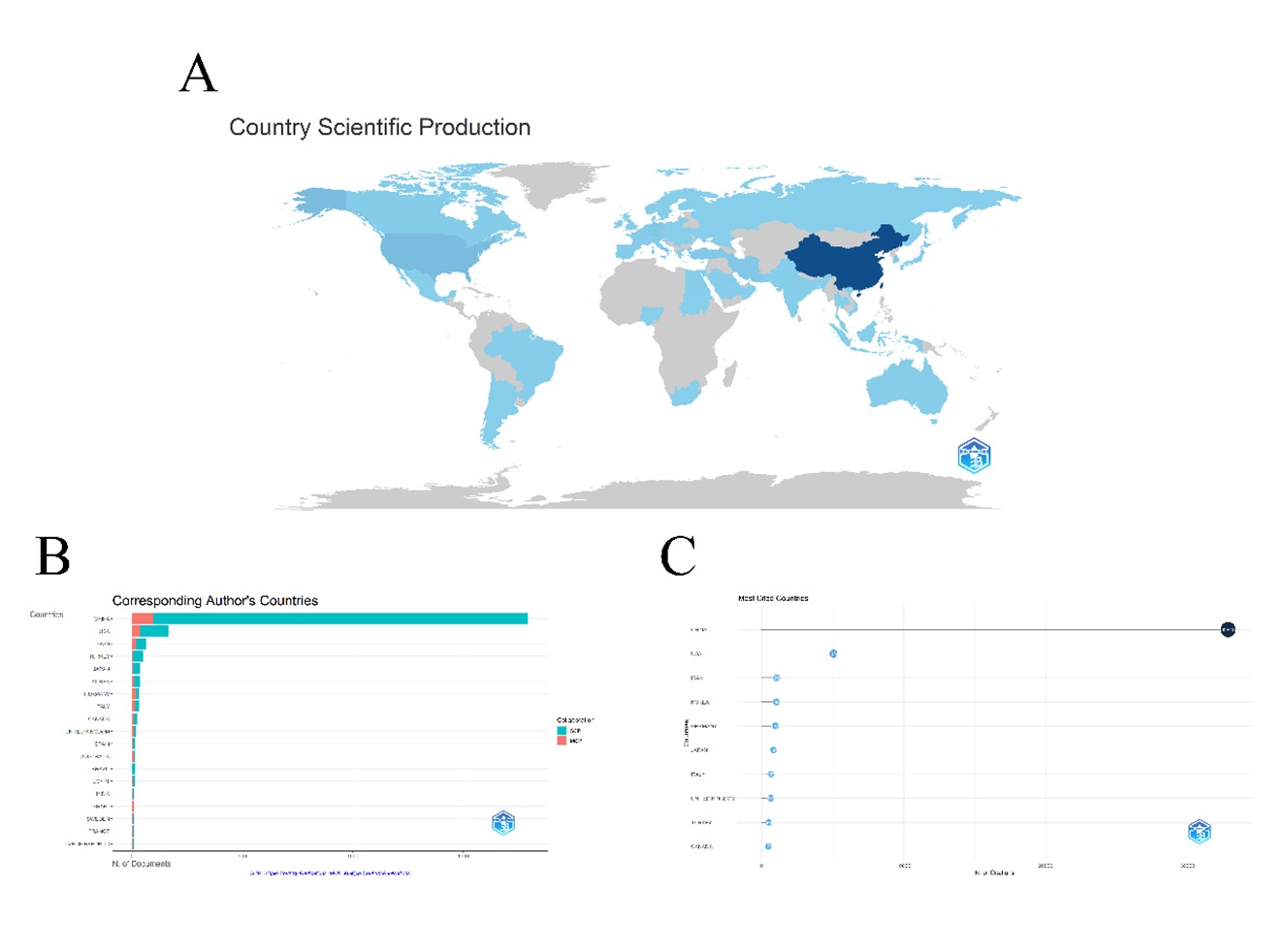
In the national scientific output map reflects the situation of the number of publications of each country and region, the gray zone in the map represents no scientific output, the darker the blue color of the country or region the more publications, it is obvious that the Chinese region is the most notable, it is the darkest color, and the number of publications is also the most, followed by the United States (Figure 6A). We then analyzed the corresponding authors from each country, and it is clear in (Figure 6B) that the corresponding authors from China far outnumber the rest of the countries, and are even more than the sum of them. It is worth noting that although most of the corresponding authors are from China, only a few of them will participate in international collaborations. With such a huge advantage in the number of authors, China is unsurprisingly ranked first among the highly cited countries, and is more than six times the size of the United States, which is ranked second.
3.6. Keywords
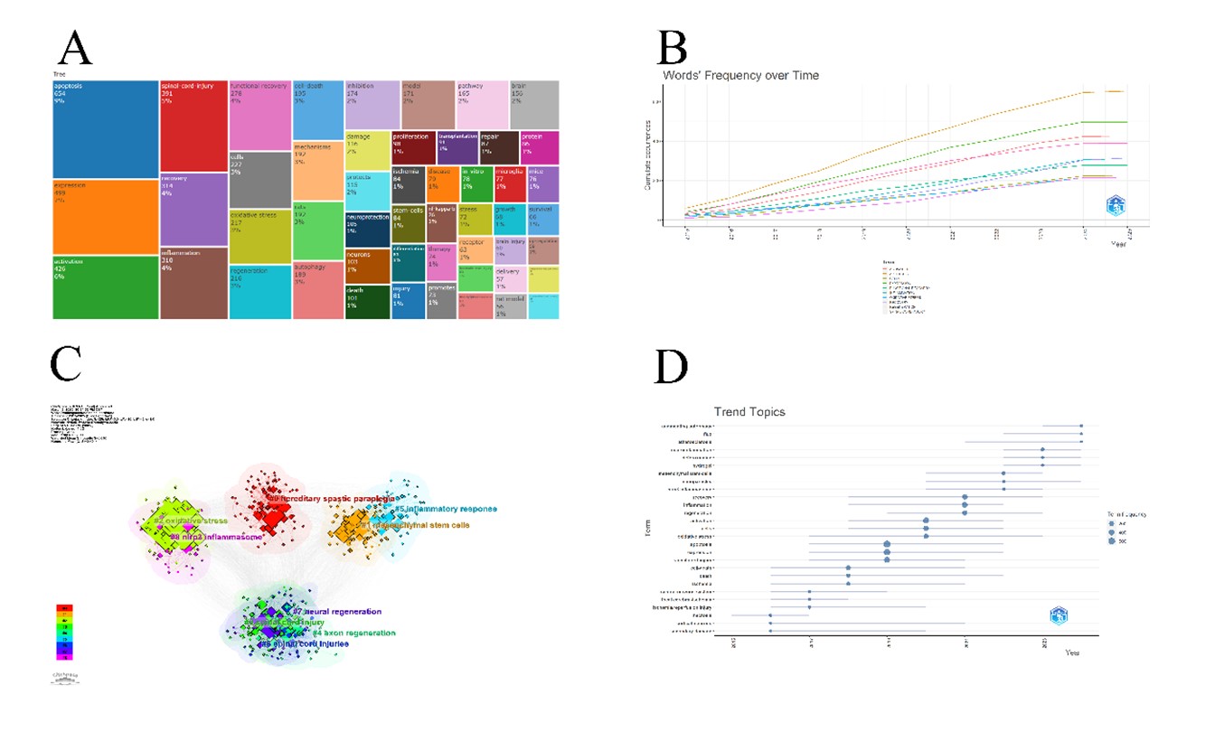
The keyword dendrogram is arranged according to the percentage of keywords, which can be summarized as follows: i) high-frequency keywords, such as “apoptosis”, “expression”, “activation”, “spinal cord injury”. These keywords accounted for ≥5% of the total; ii) medium-frequency keywords, such as “recovery”, “inflammation”, “functional recovery”, “cells”, “oxidative stress”, “regeneration”, “cell death”, ” mechanisms”, ‘rats’, ‘autophagy’, ‘inhibition’, ‘model’, ‘pathway’, ‘brain’, ‘damage’, ‘protects’. These keywords account for 2%-4%. iii) low-frequency keywords, such as “neuroprotection”, “neurons”, “proliferation”, “transplantation”, “ischemia”, “repair”, and so on. These keywords accounted for ≤1% (Figure 7A). In (Figure 7B), we can observe the changes of the top ten keywords over time. These ten keywords show a steady upward trend, with “apoptosis”, “expression”, “spinal cord injury” and “activation” dominating the list for a long time. Using CiteSpace, these articles were clustered using keywords as clues to obtain (Figure 7C). It can be seen that a total of 9 clusters were grouped.
Of these 9 clusters, (iii) and (ix) are closely related, and (iv), (v), (vii) and (viii) are closely related, and each of them can be merged into a single cluster. The new result is: i) hereditary spastic paraplegia, ii) mesenchymal stem cells, iii) nlrp3 inflammasome and oxidative stress, iv) inflammatory response, v) axon regeneration and neural regeneration.
In Trending Topics (Figure 7D) one can see the main research topics over the years and the duration of the topic. It can be seen that in terms of frequency, the keywords “expression”, “apoptosis”, and “spinal cord injury” match the results in (Figure 7A), and they are mainly active around 2019. Most of the topics ended before 2024, and a few emerging topics continued until 2024, such as “augmenting autophagy”, “flux”, “atherosclerosis”, “neuroinflammation”, “deferoxamine”, “hydrogel” and “nanoparticles”. These topics hint at possible research trends in the short term future.
3.7. Literature and Reference
The top ten local highly cited articles are displayed in (Figure 8A). The top ranked article is an original article published in Cell Death & Disease by S Liu, C Sarkar et al. in 2015, in which the authors used a rat model of contusive SCI and observed post-traumatic outcomes such as LC3-II-positive autophagosomes and accumulation of autophagy substrate protein p62 , the authors concluded that SCI leads to lysosomal impairment, which in turn leads to autophagy disruption and associated endoplasmic reticulum stress-induced neuronal apoptosis [8]. This article has been cited 86 times locally and 180 times globally, suggesting that this article not only has the highest impact locally, but also in other fields. Second on the list is an article published in Neural Regeneration Research in 2019 by Xue Yao, Yan Zhang et al. In this study, the authors conducted controlled experiments on rats to investigate the role of iron death in SCI and the therapeutic effects of deferoxamine. Their experiments showed that preoperative administration of deferoxamine reduced iron ion concentrations, attenuated iron death and gliosis, and revealed that inhibiting iron death may be a new approach to treating SCI [9]. The article was cited 58 times locally and 213 times globally, with a greater global than local impact. Ranked third is an article by Haosen Zhao, Shurui Chen et al. published in Journal of Neuroscience in 2017. The article mentions the neuroprotective effects and mechanisms of resveratrol on SCI, and their experimental results show that resveratrol treatment significantly improves motor function and reduces motor neuron loss and lesion areas in rats with SCI, and explains the mechanisms [10]. This article has 56 local citations and 167 global citations. Information about the remaining 7 articles in the ranking is in (Table 5). Figure 8B shows the global highly cited articles, and the second most highly cited local article just mentioned is ranked eighth here. This list elaborates on the global impact under this field. The first place is taken by a review published in Toxicological Sciences by Akshata Moghe, Smita Ghare et al. in 2015. The review describes the current understanding of the pathogenic mechanisms of acrolein, some of which are associated with SCI, and highlights the known and expected clinical disease effects and potential treatments [11]. The article had a total of 369 citations, underscoring its global impact. In second place was an article by Helen M. Bramlett and W. Dalton Dietrich published in 2015 in the Journal of Neurotrauma, which reviewed many pathologic mechanisms, including excitotoxicity, apoptosis, and others, exploring their consequences for traumatic brain injury, and was cited a total of 315 times [12]. Third is an article by Gregory A. Wyant, Monther Abu-Remaileh et al. published in science in 2018, in which the authors performed quantitative proteomic analysis of rapidly isolated lysosomes and identified NUFIP1 as a receptor for ribosome-selective autophagy [13]. This article was cited 258 times. The remaining seven articles on the same list are displayed in (Table 6). The references are cited in (Figure 8C), which shows the top ten references cited. The most cited article is one published in the Journal of Neurotrauma in 2009 in which authors D. Michele Basso, Michael S. Beattle and Jacqueline C. Bresnahan constructed a new motor rating scale that bears their name. This rating scale is a validated index of motor recovery, and it is predictive, distinguishing between behavioral outcomes resulting from different injuries, and predicting deconvolutional changes at the center of the injury [14]. Second place was published in Nature Reviews Disease Primers by Christopher S. Ahuja, Jefferson R. Wilson et al. This article details traumatic SCI, including pathomechanisms, animal model experiments, diagnosis and treatment [15]. Third place went to an article by D. Michele Basso, Lesley C. Fisher, et al. in the Journal of Neurotrauma, which proposed a new exercise scale (Basso Mouse Exercise Scale) that is more reliable than previously used rating scales [16]. Information on the remaining seven references is presented in (Table 7).
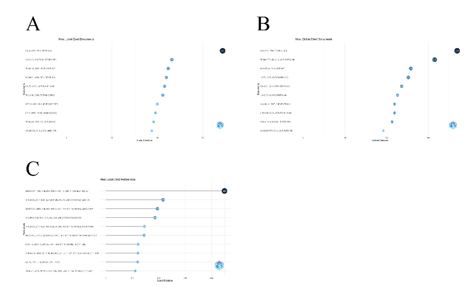
Table. 5. Top 10 locally cited articles in the target
literature.
|
Document |
DOI |
Year |
Local Citations |
Global Citations |
LC/GC Ratio (%) |
Normalized Local Citations |
Normalized Global Citations |
|
LIU S, 2015, CELL DEATH DIS |
10.1038/cddis.2014.527 |
2015 |
86 |
180 |
47.78 |
17.18 |
5.48 |
|
YAO X, 2019, NEURAL REGEN RES |
10.4103/1673-5374.245480 |
2019 |
58 |
213 |
27.23 |
11.87 |
7.32 |
|
ZHAO HS, 2017, NEUROSCIENCE |
10.1016/j.neuroscience.2017.02.027 |
2017 |
56 |
167 |
33.53 |
12.12 |
5.90 |
|
RONG YL, 2019, CELL DEATH DIS |
10.1038/s41419-019-1571-8 |
2019 |
55 |
236 |
23.31 |
11.26 |
8.11 |
|
LIU ZY, 2020, J NEUROINFLAMM |
10.1186/s12974-020-01751-2 |
2020 |
54 |
252 |
21.43 |
14.41 |
10.19 |
|
ZHOU KL, 2020, THERANOSTICS |
10.7150/thno.46566 |
2020 |
53 |
105 |
50.48 |
14.14 |
4.25 |
|
GE MH, 2021, CNS NEUROSCI THER |
10.1111/cns.13657 |
2021 |
50 |
158 |
31.65 |
14.17 |
7.86 |
|
HE M, 2016, P NATL ACAD SCI USA |
10.1073/pnas.1611282113 |
2016 |
49 |
150 |
32.67 |
10.94 |
5.66 |
|
ZHANG D, 2017, MOL NEUROBIOL |
10.1007/s12035-016-9895-1 |
2017 |
48 |
107 |
44.86 |
10.39 |
3.78 |
|
WANG CG, 2018, J CELL MOL MED |
10.1111/jcmm.13368 |
2018 |
47 |
143 |
32.87 |
11.95 |
4.77 |
Table. 6. Top 10 cited articles globally in the target
literature.
|
Paper |
DOI |
Total Citations |
TC per Year |
Normalized TC |
|
MOGHE A, 2015, TOXICOL SCI |
10.1093/toxsci/kfu233 |
369 |
33.55 |
11.23 |
|
BRAMLETT HM, 2015, J NEUROTRAUM |
10.1089/neu.2014.3352 |
315 |
28.64 |
9.59 |
|
WYANT GA, 2018, SCIENCE |
10.1126/science.aar2663 |
258 |
32.25 |
8.61 |
|
LIU ZY, 2020, J NEUROINFLAMM |
10.1186/s12974-020-01751-2 |
252 |
42.00 |
10.19 |
|
RONG YL, 2019, CELL DEATH DIS |
10.1038/s41419-019-1571-8 |
236 |
33.71 |
8.11 |
|
LIU W, 2019, J NEUROTRAUM |
10.1089/neu.2018.5835 |
226 |
32.29 |
7.76 |
|
HUANG JH, 2017, J NEUROTRAUM |
10.1089/neu.2017.5063 |
219 |
24.33 |
7.73 |
|
CHEN SB, 2018, J NEUROINFLAMM |
10.1186/s12974-018-1193-6 |
219 |
27.38 |
7.30 |
|
YAO X, 2019, NEURAL REGEN RES |
10.4103/1673-5374.245480 |
213 |
30.43 |
7.32 |
|
VACCARI JPD, 2016, J NEUROCHEM |
10.1111/jnc.13036 |
192 |
19.20 |
7.25 |
Table. 7. Top 10 cited references.
|
Cited References |
Citations |
|
BASSO DM, 1995, J NEUROTRAUM, V12, P1, DOI
10.1089/NEU.1995.12.1 |
449 |
|
AHUJA CS, 2017, NAT REV DIS PRIMERS, V3, DOI
10.1038/NRDP.2017.18 |
216 |
|
BASSO DM, 2006, J NEUROTRAUM, V23, P635, DOI
10.1089/NEU.2006.23.635 |
195 |
|
ANJUM A, 2020, INT J MOL SCI, V21, DOI
10.3390/IJMS21207533 |
186 |
|
AHUJA CS, 2017, NEUROSURGERY, V80, PS9, DOI
10.1093/NEUROS/NYW080 |
146 |
|
MCDONALD JW, 2002, LANCET, V359, P417, DOI
10.1016/S0140-6736(02)07603-1 |
145 |
|
LIVAK KJ, 2001, METHODS, V25, P402, DOI
10.1006/METH.2001.1262 |
122 |
|
OYINBO CA, 2011, ACTA NEUROBIOL EXP, V71,
P281, DOI 10.55782/ANE-2011-1848 |
121 |
|
LIU XZ, 1997, J NEUROSCI, V17, P5395 |
120 |
|
TRAN AP, 2018, PHYSIOL REV, V98, P881, DOI
10.1152/PHYSREV.00017.2017 |
111 |
4. Discussion
4.1. Global Background
SCI is a highly disabling disease of the central nervous system. It is mainly triggered by traffic accidents, falls, and diseases (e.g., tumors), and has a high prevalence in young and middle-aged males, and is accompanied by serious complications, such as urinary tract infections and respiratory dysfunction, which result in a decline in the quality of life of the patients and an increase in the burden of healthcare. In recent years, studies have shown that programmed cell death (e.g., cellular pyroptosis and necrotic apoptosis) in secondary injuries is a central mechanism that exacerbates neuroinflammation and neuronal loss. However, existing studies are mostly limited to animal experiments, and clinical translation is challenged by mechanistic complexity (e.g., the role of the non-classical pathway of pyroptosis, caspase-11), multi-pathway interactions, and the lack of humanized models.
4.2. Knowledge Base
4.2.1. Overview
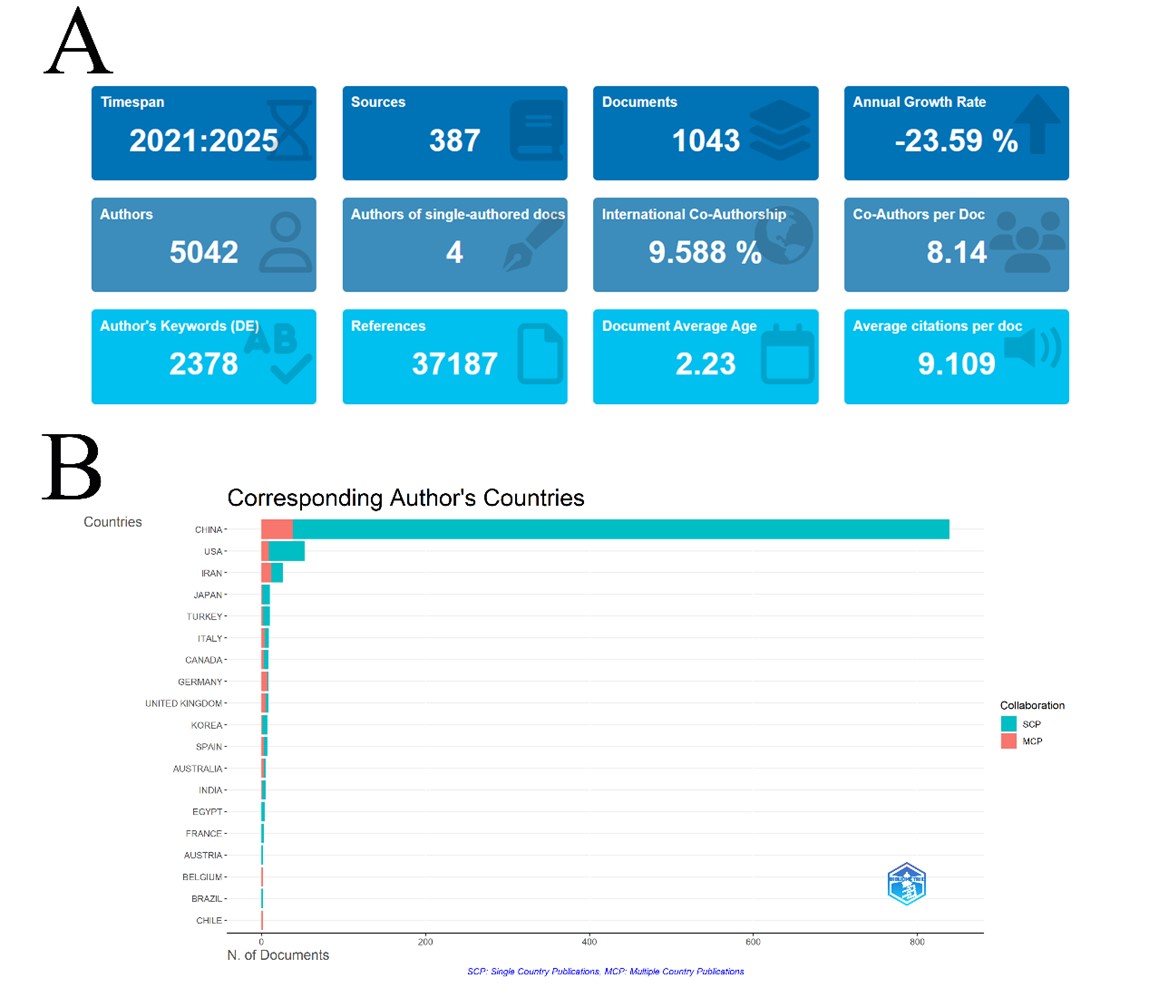
From the overview of the field we know that the scientific output of this field has slowly increased in the last few years, but the average citation frequency has decreased dramatically, and the average age of the literature during this decade is 4.95 years, suggesting that papers published in the last few years predominate. We conducted another bibliometric analysis of the literature published in the last five years, and the search rules remained the same as before. The results are shown in (Figure 9A). Since 2025 is not yet over, which leads to a large bias in the annual growth rate, this data should be ignored. We discuss a few of the more valuable data: the co-Authors per document increased from 7.62 to 8.14, suggesting that research in this area is increasingly relying on teamwork, but international co-authorship decreased to 9.588% compared to the previous 10.99%, suggesting that the authors' international partnerships is weakening. The sources of corresponding authors for the last five years show that China is still the dominant country (Figure 9B). These changes in author-related data are likely to be due to changes within China. Average citation per document has declined from 19.62 to 9.109. The large drop in average citation frequency suggests that fewer landmark studies have been published in recent years.
4.2.2. Source
In terms of journals, Molecular Neurobiology with a 2023 impact factor of 4.6 and a 5-year journal impact factor of 4.7, is categorized as Q1 in the JCR subject category NEUROSCIENCE. This journal focuses on contemporary brain research, and its main contributions include the exploration of neurobiology at the molecular level, the study of neurodegenerative diseases, and neuroinflammation and immune response, etc. It has not only made outstanding contributions to the basic research of neurobiology, but also promoted the integration of interdisciplinary research, and facilitated the application of some new technologies. Neural Regeneration Research is a journal of the Chinese Society of Rehabilitation Medicine and aims to provide timely coverage of prospective,, creative, and prevalent basic and clinical research in the field of international neuroregeneration. It has a journal impact factor of 5.9 in 2023, a five-year journal impact factor of 5.2, and is categorized as Q2 in the JCR subject category CELL BIOLOGY and Q1 in NEUROSCIENCES. This journal provides a high-quality platform for nerve regeneration research and promotes innovation in basic research, clinical translation, therapeutic approaches, and the application of new technologies for injury repair, which has a significant impact on the development of neuroscience and neuromedicine. Journal of Neurotrauma focuses on traumatic brain and spinal cord injuries and covers basic research, clinical research, treatment strategies, and mechanisms involved in neurological injuries. It has a reported impact factor of 3.9 in the 2023 Science Citation Report and a five-year status impact factor of 6.1, and is categorized into 3 JCR subject categories, CLINICAL NEUROLOGY (Q1), CRITICAL CARE MEDICINE (Q1), and NEUROSCIENCES (Q2). The difference between its 2023 impact factor and the five-year journal impact factor is 2.2, which suggests a decline in the quality of papers published in this journal. Nonetheless, it continues to make important contributions to basic research in neurotrauma and has had a significant impact on clinical translation, functional recovery, and therapeutic strategies for neurologic injuries. Journal of Neuroscience is one of the most influential and widely recognized journals in the field of neuroscience, focusing on basic and applied research on the nervous system. This journal publishes articles covering all aspects of neuroscience, including neurobiology, neurophysiology, neuropathology, and more. It has a reported impact factor of 4.4 in the 2023 Science Citation Report, a five-year journal impact factor of 5.3, and a JCR subject category of NEUROSCIENCES (Q1). Experimental Neurology advances neuroscience and its clinical care by focusing on the latest research in the areas of nerve injury, nerve repair, and neurodegenerative diseases. The journal's impact factor is reported as 4.6 in the 2023 Journal Citation Report and 4.8 for the five-year journal impact factor. The JCR subject category is NEUROSCIENCES (Q1).
4.2.3. Author
Due to the extreme distribution of authors based on the number of publications, we identified the details of only one author in the ranking of the top ten publications and the top ten cited frequencies. Jian Xiao is from China and is affiliated with Wenzhou Medical University, Chonnam National University and other institutions. He is listed in WoS under Cell Biology Pharmacology & Pharmacy Biochemistry & Molecular Biology Research & Experimental Medicine Engineering. This author's most cited article in the target literature was a review on the role and therapeutic implications of pyroptosis in SCI, which reviewed the potential role of pyroptosis-regulated programmed cell death in SCI and emphasized the targeting of pyroptosis signaling pathways and inflammatory vesicle components and their therapeutic significance in SCI [17]. Another highly cited article explored the role of endogenous FGF10 in the recovery process after neurological injury. Their study found that FGF10 is mainly produced by neurons and microglia/macrophages, and that FGF10 improves repair function after injury by reducing apoptosis as well as the FGFR2/PI3K/Akt pathway, and that inducing FGF10 expression may be a potential therapeutic lead for CNS injury [18].
4.2.4. Institution
All of the top 10 institutions in terms of publications are from China, with Wenzhou Medical University ranking first. In addition to the two highly cited papers contributed by Jian Xiao, there are several articles that have received a high number of citations, such as: A study published in 2019 on microvascular endothelial phagocytosis of myelin debris after nerve injury to inhibit inflammation and promote angiogenesis after SCI received 188 citations [19]. A 2018 publication identifying that rhodiola rosea glycosides promote functional recovery in rats with SCI and exploring the possible mechanisms involved received 158 citations [20]. A study on promoting nerve growth factor regeneration on SCI by constructing a novel heparin-poloxam thermosensitive hydrogel published in 2016 received 117 citations [21]. An article published in 2020 on the role of TFE3 in regulating autophagy after SCI received 111 citations [22]. In the seven outbreaks of institutions that have continued to date, we looked for possible reasons behind them.
i) Nanchang University: Nanchang University has a burst citation period of 2021-2025, suggesting that relatively influential articles were published before 2021. Based on this clue, we searched in WoSCC, and the result was that in 2017, Nanchang University participated in publishing a study on the molecular mechanism of the neurotrophic effects exerted by sodium valproate, and suggested that sodium valproate could be a new treatment for neurological diseases [23]; 2020 published an article on acupuncture combined with moxibustion can promote recovery from SCI [24]. These two articles have been cited 39 and 11 times respectively as of today. We consider these two articles to be the main starting cause of the citation explosion in SCSU 2021. In 2021-2025, SSC published a total of 20 articles, the citation frequency is greater than the average frequency of citations per article (9.109) in the field in the last five years, there are three articles, respectively, published in 2022 on the neuroprotective effect of Aucubin [25]. Study of the Therapeutic Effects of Alpinetin on Neuroinflammation and Neuronal Apoptosis, published in 2023 [26], Study on the Inhibition of Neuronal Iron Death after SCI by Metformin and its Relationship to Heme Oxygenase-1, published in 2024 [27].
ii) Southeast University: Southeast University leads to citation explosion starting in 2022 with co-authored review of the role of focal death in SCI [17].
iii) Anhui Medical University: The citation explosion of this institution is from 2022-2025, and we looked for the reasons for the start of its explosion, combining the citation frequency of articles published by this institution before 2022 and the time of citation, we believe that there are 3 articles that may be the main reasons for the start of this explosion of citations, they are: published 2018 in Neuroprotective effects of endothelial progenitor cells after SCI [28]. Mechanisms of microRNA-200a in bone marrow mesenchymal stem cells for SCI repair published in 2020 [29]. Molecular mechanisms of neuroprotection by erythropoietin in rats with SCI published in 2020 [30]. During the citation explosion, there were three papers with a citation frequency greater than 9.109, which may have been the main reason for supporting the explosion phenomenon in the latter years, which were: Published 2022 Studies on the Mechanism of Pericyte to Fibroblast Transformation after SCI and the Blocking Effect of Imatinib on the Transformation [31] and the molecular mechanism of iron death in secondary SCI [32]. Published 2023 Mechanisms of erythropoietin action against iron death after SCI [32].
iv) University of Science Technology of China: The institution has only 10 publications in this field, the earliest being a study published in 2020 on the protective effects of Se-CQDs after traumatic SCI in rats [33]. That article garnered 71 citations and was the start of an explosion of citations. Another study of the neuroprotective mechanisms of Quietness Circ 0000962 in the same year garnered 22 citations and also set the stage for an outbreak of citation [34]. After this, there are three other articles with citation frequencies greater than 9.109, two experiments on the development of genetically engineered MicroRNA 21-loaded exosomes for the repair of spinal cord injuries [35] and a study on nanomaterial-assisted siRNA silencing of IRF5 to regulate macrophage polarization for SCI repair, both published in 2022 [36]; 2023 Publication on Exploration of Mechanisms of Microelectric Field-Induced Nerve Injury Repair and Related Potential Therapeutic Strategies [37].
v) Jinan University: This institution published a total of 25 relevant articles, with several having a total citation frequency greater than the field average. It has a burst citation start year of 2023, in which three articles had a sudden increase in citation frequency that became the main reason for the start of the burst citation, they are: research on designing multifunctional selenium nanoparticles to attenuate SCI induced by oxidative stress, published in 2019 [38]. Injectable hydrogels to control curcumin release to improve the microenvironment for SCI repair, published in 2021 [39], and selenium nanoparticles piggybacking on herbal ingredients to increase the efficiency of herbal medicine use for the purpose of treating SCI, published in 2022 [40]. A study published in 2023 on the construction of nanoenzymes loaded with rapamycin and hollow mesoporous Prussian blue for combined antioxidant and anti-inflammatory treatment of SCI received 17 citations in 2024 [41]. These four articles, along with others, form a continuation of the burst of citations.
vi) Tsinghua University: This institution had 14 publications, and a study published in 2021 on highly permeable DNA supramolecular hydrogels to promote recovery after SCI became the main reason for the citation burst in 2023 [42]. An exploration of nanoscaffolds combined with stem cell transplantation therapeutic strategies published in 2021 and a study on the promotion of SCI repair through the antioxidant effect of single-atom cobalt nano-enzymes and by constructing a double-crosslinked biomimetic composite hydrogel piggybacked with WAY-316606 for inducing repair after SCI published in 2023 [43, 44], together with other articles, form a continuation of the citation burst phenomenon.
vii) Huzhou University: According to the WoS citation report, the main citation burst at Huzhou University occurred in 2023, a year in which a study on the therapeutic mechanism of betulinic acid on SCI published in 2021 received 31 citations and became the main reason for this burst phenomenon [45]. This subsequent article was also cited at a high frequency, continuing the outbreak phenomenon.
4.2.5. Country or Region
China has an all-around lead in this field of research, with the vast majority of authors from China, but most of them are not connected to the international community. Looking at the authors' collaborative networks we can easily see that the collaborative relationships between authors show clear subgroups. And there are few collaborative relationships between groups and groups, and between in-group members and out-group members. We hypothesize that most of the researchers from China use intra-group cooperation as their main mode of cooperation. They are neither good at collaborating on research with people from other groups, nor are they good at collaborating with international researchers. Combined with the decreasing average citation frequency, we further hypothesize that most of these groups undertake simpler research tasks. These tasks can be accomplished with help from within the group alone, which may explain why authors from China possess a lower rate of international collaboration. Analyzing the distribution of authors by number of publications, we learned that most authors in this field have very few publications, which may indicate that there is a gradual influx of new authors into this field. One possible reason for this phenomenon is the expansion of graduate school enrollment in Chinese medical education institutions. It is recommended that the Chinese government and institutions encourage researchers to engage in broader exchanges and collaborations to facilitate the contribution of scholarly achievements and the transfer of key technologies. Encourage more valuable research and improve the average citation frequency, which is declining year by year.
4.2.6. Keyword
The high-frequency keywords “apoptosis”, “expression”, “activation”, “SCI” highlight the research hotspots in this field. the programmed death that occurs after SCI is a key process in the deterioration of this disease, and whether it can be blocked or inhibited will directly affect the treatment effect. Current research has found multiple ways to cope with different types of apoptosis, such as induction of FGF10 expression to reduce apoptosis, as mentioned above, and the use of iron death inhibitors or erythropoietin to inhibit iron death and promote SCI repair [9]. Endoplasmic reticulum stress after SCI induces neuronal apoptosis, and preventing endoplasmic reticulum stress may also be a therapeutic approach.
4.3. Keyword Clustering Results
4.3.1. Hereditary Spastic Paraplegia
This cluster focuses on hereditary spastic paraplegia, which is characterized by progressively worsening spasticity and weakness of the lower extremities, often associated with some lesion of the corticospinal tract. Although it is a genetic disease, its molecular mechanisms and neurodegenerative processes have some commonalities with axonal damage and regenerative disorders following SCI, so the theoretical basis of SCI is drawn upon in these studies or SCI is introduced as a control [46-48].
4.3.2. Mesenchymal Stem Cells
This cluster focuses on the application of mesenchymal stem cells (MSCs) in SCI repair. MSCs are highly regarded in regenerative medicine due to their multidirectional differentiation ability, immunomodulatory effects and secretion of neurotrophic factors. Related research directions include: Cell transplantation and tissue engineering: using MSC to fill the injured area and improve the local microenvironment. Immunomodulation and anti-inflammatory effects: regulating post-injury inflammatory responses and promoting neuroprotection by secreting cytokines. Promoting nerve regeneration: MSC help activate endogenous repair mechanisms and rebuild neural networks [49-51].
4.3.3. NLRP3 inflammasome and Oxidative Stress
This cluster focuses on the secondary pathological processes following SCI, particularly the inflammatory response and oxidative stress. Activation of the NLRP3 inflammatory vesicle, an important molecular complex that regulates the inflammatory response, leads to a massive release of pro-inflammatory cytokines that exacerbate neuronal injury. Post-injury reactive oxygen species (ROS) production and its destruction of cellular structures are equally important mechanisms of secondary injury. The aim of this line of research is to reveal these molecular mechanisms and explore intervention strategies to inhibit NLRP3 activation or alleviate oxidative stress, thereby reducing secondary injury and improving the environment for neural repair [52-54].
4.3.4. Inflammatory Response
This cluster is broader than the previous one and covers the study of the overall inflammatory response after SCI. The main components include: inflammatory cell activation and infiltration: e.g., activation of microglia and macrophages, M1/M2 polarization, and cytokines secreted by them; inflammatory mediators and signaling pathways: e.g., the role of cytokines, such as TNF-α, IL-1β, and IL-6, and their regulatory mechanisms. The research in this direction aims to comprehensively understand how the inflammatory response may both exacerbate injury and provide necessary signals for repair, thus providing theoretical basis and therapeutic targets for clinical regulation of the inflammatory response to promote recovery [55-57].
4.3.5. Axon Regeneration and Neural Regeneration
This is the most central direction in SCI repair research, focusing on promoting regeneration and reconstruction of damaged nerves. The main content includes: axon regeneration mechanism: how to overcome the inhibitory environment (e.g. glial scar, inhibitory molecules) to promote axon extension; neural network reconstruction: not only focus on the regeneration of single axon, but also aim to rebuild the complete nerve conduction pathway and functional neural network; this direction of research is the key to realize the functional restoration, which is directly related to the reconstruction of motor and sensory functions of the patients with spinal cord injuries [58-60].
4.4. Research Trends
4.4.1. Modulation of Autophagic Flux to Promote Recovery from SCI
Cellular autophagy plays a key role in SCI, usually after SCI, cells regulate intracellular protein structure through autophagy, and appropriate autophagic flux contributes to the repair of neuronal cell injury. A number of studies have been conducted to determine the positive effects of regulating autophagic flux on the repair of neuronal cell injury. Subsequent studies should continue to explore the underlying mechanisms and regulation of this process [61, 62].
4.4.2. Mechanisms of Atherosclerosis-Like Responses after SCI and Ways to Block Them
After SCI, ischemia and hypoxia at the site of injury are important causes of subsequent exacerbations. It has been shown that a variety of ischemia- and hypoxia-related differentially expressed genes are significantly expressed after SCI, and most of these genes are enriched in atherosclerosis-related pathways [63]. Not only that, but the formation of foam cells after phagocytosis of myelin debris by macrophages after SCI is also similar to the formation of atherosclerosis [64]. Subsequent studies should continue to explore related pathogenic mechanisms and interventions.
4.4.3. Exploratory and Interventional Approaches to Inflammatory Mechanisms after SCI
Inflammation is an important component of SCI and involves a wide range of factors, such as immune cells, the surrounding microenvironment, and related gene expression. Activation of microglia and astrocytes promotes the release of inflammatory factors and the production of reactive oxygen species, which exacerbate neuronal damage. It has been shown that the release of inflammatory factors can be inhibited or the microglia phenotype can be modulated by certain means [65, 66]. In the future, it will be necessary to explore possible mechanisms and to translate what has been discovered into the clinic.
4.4.4. Inhibition of Iron Death in SCI and the Use of Desferrioxamine
Desferrioxamine is a chelating agent that has been found in recent years to have multiple effects in nerve injury repair, including chelation of free iron ions to reduce iron-dependent cell death and down-regulation of NLRP3 inflammatory vesicles to reduce ROS levels. Many studies have affirmed the therapeutic effects of deferoxamine in SCI [67, 68]. Subsequent clinical trials are needed to optimize the use and mode of administration as well as to determine adverse effects.
4.4.5. Development of Drug Carriers
A variety of drugs have been identified as having therapeutic effects on spinal cord injuries, but due to the unique properties of some of these drugs and the specificity of the injury site, these drugs are difficult to act directly on the injury site, and a carrier is required to transport the drugs [69-71]. In the future, more carrier-drug combinations can be explored to accelerate the translation of therapeutic programs to the clinic.
5. Conclusion
In this study, we carried out a bibliometric and visualization analysis of the last decade of research on spinal cord injuries associated with programmed death. We determined the overall picture of the field, including changes in the number of publications and average citation frequency per year, listed the core journals under this field, identified the high contributing authors, institutions, and countries under this field. We discuss in detail the contributions of institutions and the special situations that have arisen, and make predictions about future research trends based on trending topics.
Limitation
In this paper, we bibliometric and visualize the literature in the target domain through bibliometric methods, and this study has some limitations due to the specificity of the type: i) The purpose of this type of study is to mine the existing literature and mine information from multiple perspectives in the target domain to construct a knowledge graph, but not to propose specific treatment options or other insights. ii) The data were collected from the WoSCC, Scopus and pubmed. We affirmed the quality of the literature that was included in them, and the literature that was not included in them was not included in this study. This may have caused some bias, but this bias was minor and did not affect the main results; iii) We conducted a study of the literature in the field over the last ten years, and the results obtained reflect the state of the art over the ten-year period; literature after the cutoff date was not included, which means that the results we obtained have a certain degree of timeliness.
Ethics Approval
None.
Author Contributions
XY Wang and T Liu contributed equally to this work. ZQ Hou, SH Xie, XY Wang and T Liu were involved in the conceptualization of the study, the analysis of the results and the drafting of the manuscript. XY Wang, Z Zhang and YF Jiang conducted a systematic literature search, Z Zhang and YF Jiang independently screened the target literature and compared the differences. XY Wang determined the final results in case of disagreement. XY Wang and YF Jiang jointly processed the data, and T Liu critiqued the content and methodology of the study.
Data Availability Statement
The data that support the findings of this study are available from the corresponding author upon reasonable request.
Acknowledgments
None.
Funding
None.
Conflicts of Interest
2.3. Statistical Analysis
None.
REFERENCES
[1] John W McDonald, Cristina
Sadowsky “Spinal-cord injury.” Lancet, vol. 359, no. 9304, pp. 417-425,
2002. View at: Publisher
Site | PubMed
[2] Guangjin Gu, Huaqi Yu,
Huishuang Zou, et al. “Pyroptosis in Spinal Cord Injury: Implications for
Pathogenesis and Therapeutic Approaches.” Front Biosci (Landmark Ed),
vol. 29, no. 6, pp. 210, 2024. View at: Publisher Site | PubMed
[3] Elizabeth M Kenny, Emin
Fidan, Qin Yang, et al. “Ferroptosis contributes to neuronal death and
functional outcome after traumatic brain injury.” Crit Care Med, vol.
47, no. 3, pp. 410-418, 2019. View at: Publisher Site | PubMed
[4] Chaochen Li, Chunshuai Wu,
Chunyan Ji, et al. “The pathogenesis of DLD-mediated cuproptosis induced spinal
cord injury and its regulation on immune microenvironment.” Front Cell
Neurosci, vol. 17, pp. 1132015, 2023. View at: Publisher Site | PubMed
[5] Jun-Bo Tu, Tao Liu,
Jun-Feng Li, et al. “Global research trends and hotspots in metabolomics of
osteosarcoma: a decade-spanning bibliometric and visualized analysis.” Front
Immunol, vol. 15, pp. 1463078, 2024. View at: Publisher Site | PubMed
[6] Chaomei Chen “CiteSpace
II: Detecting and visualizing emerging trends and transient patterns in
scientific literature.” Journal of the American Society for Information
Science and Technology, vol. 57, no. 3, pp. 359-377, 2005. View at: Publisher Site
[7] Massimo Aria, Corrado
Cuccurullo “bibliometrix: An R-tool for comprehensive science mapping
analysis.” Journal of informetrics, vol. 11, no. 4, pp. 959-975, 2017.
View at: Publisher
Site
[8] S Liu, C Sarkar, M Dinizo,
et al. “Disrupted autophagy after spinal cord injury is associated with ER
stress and neuronal cell death.” Cell Death Dis, vol. 6, no. 1, pp.
e1582, 2015. View at: Publisher
Site | PubMed
[9] Xue Yao, Yan Zhang, Jian
Hao, et al. “Deferoxamine promotes recovery of traumatic spinal cord injury by
inhibiting ferroptosis.” Neural Regen Res, vol. 14, no. 3, pp. 532-541,
2019. View at: Publisher
Site | PubMed
[10] Haosen Zhao, Shurui Chen,
Kai Gao, et al. “Resveratrol protects against spinal cord injury by activating
autophagy and inhibiting apoptosis mediated by the SIRT1/AMPK signaling
pathway.” Neuroscience, vol. 348, pp. 241-251, 2017. View at: Publisher
Site | PubMed
[11] Akshata Moghe, Smita
Ghare, Bryan Lamoreau, et al. “Molecular mechanisms of acrolein toxicity:
relevance to human disease.” Toxicol Sci, vol. 143, no. 2, pp. 242-255,
2015. View at: Publisher
Site | PubMed
[12] Helen M Bramlett, W Dalton
Dietrich “Long-Term Consequences of Traumatic Brain Injury: Current Status of
Potential Mechanisms of Injury and Neurological Outcomes.” J Neurotrauma,
vol. 32, no. 23, pp. 1834-1848, 2015. View at: Publisher Site | PubMed
[13] Gregory A Wyant, Monther
Abu-Remaileh, Evgeni M Frenkel, et al. “NUFIP1 is a ribosome receptor for
starvation-induced ribophagy.” Science, vol. 360, no. 6390, pp. 751-758,
2018. View at: Publisher
Site | PubMed
[14] D M Basso, M S Beattie, J
C Bresnahan “A sensitive and reliable locomotor rating scale for open field
testing in rats.” J Neurotrauma, vol. 12, no. 1, pp. 1-21, 1995. View
at: Publisher Site | PubMed
[15] Christopher S Ahuja,
Jefferson R Wilson, Satoshi Nori, et al. “Traumatic spinal cord injury.” Nat
Rev Dis Primers, vol. 3, no. 1, pp. 1-21, 2017. View at: Publisher Site | PubMed
[16] D Michele Basso, Lesley C
Fisher, Aileen J Anderson, et al. “Basso Mouse Scale for locomotion detects
differences in recovery after spinal cord injury in five common mouse strains.”
J Neurotrauma, vol. 23, no. 5, pp. 635-659, 2006. View at: Publisher Site | PubMed
[17] Abdullah Al Mamun, Yanqing
Wu, Ilma Monalisa, et al. “Role of pyroptosis in spinal cord injury and its
therapeutic implications.” J Adv Res, vol. 28, pp. 97-109, 2020. View
at: Publisher
Site | PubMed
[18] Jian Chen, Zhouguang Wang,
ZengMing Zheng, et al. “Neuron and microglia/macrophage-derived FGF10 activate
neuronal FGFR2/PI3K/Akt signaling and inhibit microglia/macrophages TLR4/NF-κB-dependent
neuroinflammation to improve functional recovery after spinal cord injury.” Cell Death Dis,
vol. 8, no. 10, pp. e3090, 2017. View at: Publisher Site | PubMed
[19] Tian Zhou, Yiming Zheng,
Li Sun, et al. “Microvascular endothelial cells engulf myelin debris and
promote macrophage recruitment and fibrosis after neural injury.” Nat
Neurosci, vol. 22, no. 3, pp. 421-435, 2019. View at: Publisher Site | PubMed
[20] Chenggui Wang, Qingqing
Wang, Yiting Lou, et al. “Salidroside attenuates neuroinflammation and improves
functional recovery after spinal cord injury through microglia polarization
regulation.” J Cell Mol Med, vol. 22, no. 2, pp. 1148-1166, 2018. View
at: Publisher Site | PubMed
[21] Ying-Zheng Zhao, Xi Jiang,
Jian Xiao, et al. “Using NGF heparin-poloxamer thermosensitive hydrogels to
enhance the nerve regeneration for spinal cord injury.” Acta Biomater,
vol. 29, pp. 71-80, 2016. View at: Publisher Site | PubMed
[22] Kailiang Zhou, Zhilong
Zheng, Yao Li, et al. “TFE3, a potential therapeutic target for Spinal Cord
Injury via augmenting autophagy flux and alleviating ER stress.” Theranostics,
vol. 10, no. 20, pp. 9280, 2020. View at: Publisher Site | PubMed
[23] Zhengmao Li, Fenzan Wu,
Xie Zhang, et al. “Valproate attenuates endoplasmic reticulum stress-induced
apoptosis in SH-SY5Y cells via the AKT/GSK3β signaling pathway Int J Mol Sci, vol. 18, no. 2, pp. 315,
2017. View at: Publisher
Site | PubMed
[24] Li-Li-Qiang Ding,
Song-Feng Hu, Xing-Wei He, et al. “Acupuncture combined with moxibustion
promote the recovery of spinal cord injury in correlation with Shh/Gli-1
signaling pathway.” J Spinal Cord Med, vol. 45, no. 1, pp. 106-116,
2022. View at: Publisher
Site | PubMed
[25] Shining Xiao, Nanshan
Zhong, Quanming Yang, et al. “Aucubin promoted neuron functional recovery by
suppressing inflammation and neuronal apoptosis in a spinal cord injury model.”
International Immunopharmacology, vol. 111, pp. 109163, 2022. View at: Publisher Site | PubMed
[26] Shining Xiao, Yu Zhang,
Zihao Liu, et al. “Alpinetin inhibits neuroinflammation and neuronal apoptosis
via targeting the JAK2/STAT3 signaling pathway in spinal cord injury.” CNS
Neurosci Ther, vol. 29, no. 4, pp. 1094-1108, 2023. View at: Publisher Site | PubMed
[27] Zhihua Wang, Wu Zhou,
Zhixiong Zhang, et al. “Metformin alleviates spinal cord injury by inhibiting
nerve cell ferroptosis through upregulation of heme oxygenase-1 expression.” Neural
Regen Res, vol. 19, no. 9, pp. 2041-2049, 2024. View at: Publisher Site | PubMed
[28] Tao Wang, Xiao Fang,
Zong-Sheng Yin “Endothelial progenitor cell-conditioned medium promotes
angiogenesis and is neuroprotective after spinal cord injury.” Neural Regen
Res, vol. 13, no. 5, pp. 887-895, 2018. View at: Publisher Site | PubMed
[29] Xianxiang Wang, Lei Ye, Ke
Zhang, et al. “Upregulation of microRNA‐200a in bone marrow mesenchymal stem cells enhances the repair of spinal
cord injury in rats by reducing oxidative stress and regulating Keap1/Nrf2
pathway.” Artif Organs, vol. 44, no. 7, pp. 744-752, 2020. View
at: Publisher Site | PubMed
[30] Lin Zhong, Hui Zhang,
Zheng-Fei Ding, et al. “Erythropoietin-induced autophagy protects against
spinal cord injury and improves neurological function via the
extracellular-regulated protein kinase signaling pathway.” Mol Neurobiol,
vol. 57, no. 10, pp. 3993-4006, 2020. View at: Publisher Site | PubMed
[31] Fei Yao, Yang Luo,
Yan-Chang Liu, et al. “Imatinib inhibits pericyte-fibroblast transition and
inflammation and promotes axon regeneration by blocking the PDGF-BB/PDGFRβ
pathway in spinal cord injury.” Inflamm Regen, vol. 42, no.
1, pp. 44, 2022. View at: Publisher
Site | PubMed
[32] Yu Kang, Qiangwei Li, Rui
Zhu, et al. “Identification of ferroptotic genes in spinal cord injury at
different time points: bioinformatics and experimental validation.” Mol
Neurobiol, vol. 59, no. 9, pp. 5766-5784, 2022. View at: Publisher Site | PubMed
[33] Wenqi Luo, Yiming Wang,
Feng Lin, et al. “Selenium-doped carbon quantum dots efficiently ameliorate
secondary spinal cord injury via scavenging reactive oxygen species.” Int J
Nanomedicine, vol. 15, pp. 10113-10125, 2020. View at: Publisher Site | PubMed
[34] Rui He, Guo-Lin Tang, Lei
Niu, et al. “Quietness Circ 0000962 promoted nerve cell inflammation through
PIK3CA/Akt/NF-κB signaling by miR-302b-3p in spinal cord injury.” Ann Palliat Med,
vol. 9, no. 2, pp. 190-198, 2020. View at: Publisher Site | PubMed
[35] Xingzhi Liu, Lulu Zhang,
Zhongjuan Xu, et al. “A functionalized collagen-I scaffold delivers microRNA
21-loaded exosomes for spinal cord injury repair.” Acta Biomater, vol.
154, pp. 385-400, 2022. View at: Publisher Site | PubMed
[36] Dezun Ma, He Shen, Fangman
Chen, et al. “Inflammatory microenvironment‐responsive nanomaterials promote spinal cord injury repair by targeting
IRF5.” Adv Healthc Mater, vol. 11, no. 23, pp. 2201319,
2022. View at: Publisher
Site | PubMed
[37] Kewei Li, Zhong Liu,
Peipei Wu, et al. “Micro electrical fields induced MSC-sEVs attenuate neuronal
cell apoptosis by activating autophagy via lncRNA MALAT1/miR-22-3p/SIRT1/AMPK
axis in spinal cord injury.” J Nanobiotechnology, vol. 21, no. 1, pp.
451, 2023. View at: Publisher
Site | PubMed
[38] Siyuan Rao, Yongpeng Lin,
Yanxin Du, et al. “Designing multifunctionalized selenium nanoparticles to
reverse oxidative stress-induced spinal cord injury by attenuating ROS
overproduction and mitochondria dysfunction.” J Mater Chem B, vol. 7,
no. 16, pp. 2648-2656, 2019. View at: Publisher Site | PubMed
[39] Jinghua Luo, Xueshuang
Shi, Liming Li, et al. “An injectable and self-healing hydrogel with controlled
release of curcumin to repair spinal cord injury.” Bioact Mater, vol. 6,
no. 12, pp. 4816-4829, 2021. View at: Publisher Site | PubMed
[40] Siyuan Rao, Yongpeng Lin,
Rui Lin, et al. “Traditional Chinese medicine active ingredients-based selenium
nanoparticles regulate antioxidant selenoproteins for spinal cord injury
treatment.” J Nanobiotechnology, vol. 20, no. 1, pp. 278, 2022. View at:
Publisher Site | PubMed
[41] Kui Shen, Xiaowei Li,
Guanning Huang, et al. “High rapamycin-loaded hollow mesoporous Prussian blue
nanozyme targets lesion area of spinal cord injury to recover locomotor
function.” Biomaterials, vol. 303, pp. 122358, 2023. View at: Publisher
Site | PubMed
[42] Taoyang Yuan, Yu Shao, Xu
Zhou, et al. “Highly permeable DNA supramolecular hydrogel promotes
neurogenesis and functional recovery after completely transected spinal cord
injury.” Adv Mater, vol. 33, no. 35, pp. e2102428, 2021. View at: Publisher Site | PubMed
[43] Yuxing Jiang, Hongtao
Rong, Yifan Wang, et al. “Single-atom cobalt nanozymes promote spinal cord
injury recovery by anti-oxidation and neuroprotection.” Nano Research,
vol. 16, no. 7, pp. 9752-9759, 2023. View at: Publisher Site
[44] Xiaokun Chen, Yiyang Wang,
Gang Zhou, et al. “The combination of nanoscaffolds and stem cell
transplantation: Paving a promising road for spinal cord injury regeneration.” Biomed
Pharmacother, vol. 143, pp. 112233, 2021. View at: Publisher Site | PubMed
[45] Chenyu Wu, Huanwen Chen,
Rong Zhuang, et al. “Betulinic acid inhibits pyroptosis in spinal cord injury
by augmenting autophagy via the AMPK-mTOR-TFEB signaling pathway.” Int J
Biol Sci, vol. 17, no. 4, pp. 1138, 2021. View at: Publisher Site | PubMed
[46] Carolina Montoro-Gámez,
Hendrik Nolte, Thibaut Molinié, et al. “SARM1 deletion delays cerebellar but
not spinal cord degeneration in an enhanced mouse model of SPG7 deficiency.” Brain,
vol. 146, no. 10, pp. 4117-4131, 2023. View at: Publisher Site | PubMed
[47] Matthieu Béreau, Mathieu
Anheim, Jean-Baptiste Chanson, et al. “Dalfampridine in hereditary spastic
paraplegia: a prospective, open study.” J Neurol, vol. 262, no. 5, pp.
1285-1288. View at: Publisher
Site | PubMed
[48] Yvonne S Yang, Stephen M
Strittmatter “The reticulons: a family of proteins with diverse functions.” Genome
Biol, vol. 8, no. 12, pp. 234, 2007. View at: Publisher Site | PubMed
[49] Arnold I Caplan “Adult
mesenchymal stem cells for tissue engineering versus regenerative medicine.” J
Cell Physiol, vol. 213, no. 2, pp. 341-347, 2007. View at: Publisher Site | PubMed
[50] C P Hofstetter, E J
Schwarz, D Hess, et al. “Marrow stromal cells form guiding strands in the
injured spinal cord and promote recovery.” Proc Natl Acad Sci U S A,
vol. 99, no. 4, pp. 2199-2204, 2002. View at: Publisher Site | PubMed
[51] Yufang Shi, Yu Wang, Qing
Li, et al. “Immunoregulatory mechanisms of mesenchymal stem and stromal cells
in inflammatory diseases.” Nat Rev Nephrol, vol. 14, no. 8, pp. 493-507,
2018. View at: Publisher
Site | PubMed
[52] Yan Zhang, Wenkai Zhang,
Tao Liu, et al. “Upregulation of circ0000381 attenuates microglial/macrophage
pyroptosis after spinal cord injury.” Neural Regen Res, vol. 19, no. 6,
pp. 1360-1366, 2024. View at: Publisher
Site | PubMed
[53] Xiao Fan, Wei Ma, Yingyu
Zhang, et al. “P2X7 receptor (P2X7R) of microglia mediates neuroinflammation by
regulating (NOD)-like receptor protein 3 (NLRP3) inflammasome-dependent
inflammation after spinal cord injury.” Med Sci Monit, vol. 26, pp.
e925491, 2020. View at: Publisher
Site | PubMed
[54] Conghui Zhou, Jinfeng
Zheng, Yunpeng Fan, et al. “TI: NLRP3 inflammasome-dependent pyroptosis in CNS
trauma: a potential therapeutic target.” Front Cell Dev Biol, vol. 10,
pp. 821225, 2022. View at: Publisher
Site | PubMed
[55] Amy L Bowes, Ping K Yip
“Modulating inflammatory cell responses to spinal cord injury: all in good
time.” J Neurotrauma, vol. 31, no. 21, pp. 1753-1766, 2014. View at: Publisher Site | PubMed
[56] B Zhang, J C Gensel “Is
neuroinflammation in the injured spinal cord different than in the brain?
Examining intrinsic differences between the brain and spinal cord.” Exp
Neurol, vol. 258, pp. 112-120, 2014. View at: Publisher Site | PubMed
[57] Liqun Yang, Peter C
Blumbergs, Nigel R Jones, et al. “Early expression and cellular localization of
proinflammatory cytokines interleukin-1β, interleukin-6, and tumor
necrosis factor-α in human traumatic spinal cord
injury.” Spine, vol. 29, no. 9, pp. 966-971, 2004. View at: Publisher Site | PubMed
[58] Ken Kadoya, Paul Lu, Kenny
Nguyen, et al. “Spinal cord reconstitution with homologous neural grafts
enables robust corticospinal regeneration.” Nat Med, vol. 22, no. 5, pp.
479-487, 2016. View at: Publisher
Site | PubMed
[59] Mingyong Gao, Paul Lu,
Bridget Bednark, et al. “Templated agarose scaffolds for the support of motor
axon regeneration into sites of complete spinal cord transection.” Biomaterials,
vol. 34, no. 5, pp. 1529-1536, 2013. View at: Publisher
Site | PubMed
[60] Liya Lin, Hefeng Lin, Shi
Bai, et al. “Bone marrow mesenchymal stem cells (BMSCs) improved functional
recovery of spinal cord injury partly by promoting axonal regeneration.” Neurochem
Int, vol. 115, pp. 80-84, 2018. View at: Publisher Site | PubMed
[61] Kailiang Zhou, Charles A
Sansur, Huazi Xu, et al. “The temporal pattern, flux, and function of autophagy
in spinal cord injury.” Int J Mol Sci, vol. 18, no. 2, pp. 466, 2017.
View at: Publisher Site | PubMed
[62] Zhiheng Chen, Shengting
Wu, Shihao Sheng, et al. “hUCMSC-derived exosomes mitigate blood-spinal cord
barrier disruption by activating AMPK/mTOR-mediated autophagic flux after acute
spinal cord injury.” Composites Part B: Engineering, vol. 289, pp.
111944, 2025. View at: Publisher
Site
[63] Lijuan Zhu, Na Gao, Zhibo
Zhu, et al. “Bioinformatics analysis of differentially expressed genes related
to ischemia and hypoxia in spinal cord injury and construction of miRNA-mRNA or
mRNA-transcription factor interaction network.” Toxicol Mech Methods,
vol. 34, no. 3, pp. 300-318, 2024. View at: Publisher Site | PubMed
[64] Chenxi Zhang, Shujie Zhao,
Zhenfei Huang, et al. “Macropinocytosis enhances foamy macrophage formation and
cholesterol crystallization to activate NLRP3 inflammasome after spinal cord
injury.” Redox Biol, vol. 79, pp. 103469, 2025. View at: Publisher Site | PubMed
[65] Ziwei Fan, Mengxian Jia,
Jian Zhou, et al. “Pharmacological targeting cGAS/STING/NF-kappaB axis by
tryptanthrin induces microglia polarization toward M2 phenotype and promotes
functional recovery in a mouse model of spinal cord injury.” Neural Regen
Res, vol. 20, no. 11, pp. 3287-3301, 2025. View at: Publisher Site | PubMed
[66] Hassan Saad, Bachar El
Baba, Ali Tfaily, et al. “Complement-dependent neuroinflammation in spinal cord
injury: from pathology to therapeutic implications.” Neural Regen Res,
vol. 20, no. 5, pp. 1324-1335, 2025. View at: Publisher Site | PubMed
[67] Jian Hao, Bo Li, Hui-Quan
Duan, et al. “Mechanisms underlying the promotion of functional recovery by
deferoxamine after spinal cord injury in rats.” Neural Regen Res, vol.
12, no. 6, pp. 959-968, 2017. View at: Publisher Site | PubMed
[68] Irene Paterniti, Emanuela
Mazzon, Esposito Emanuela, et al. “Modulation of inflammatory response after
spinal cord trauma with deferoxamine, an iron chelator.” Free Radic Res,
vol. 44, no. 6, pp. 694-709, 2010. View at: Publisher Site | PubMed
[69] M Douglas Baumann,
Catherine E Kang, Charles H Tator, et al. “Intrathecal delivery of a polymeric
nanocomposite hydrogel after spinal cord injury.” Biomaterials, vol. 31,
no. 30, pp. 7631-7639, 2010. View at: Publisher
Site | PubMed
[70] Shishi He, Yanni Fu,
Zicong Tan, et al. “Optimization of ultra-small nanoparticles for enhanced drug
delivery.” BIO Integration, vol. 4, no. 2, pp. 62, 2022. View at: Publisher Site
[71] Nisha Kalayil, Aarati Anil Budar, Roshan Kalulal Dave “Nanofibers for Drug Delivery: Design and Fabrication Strategies.” BIO Integration, vol. 5, no. 1, pp. 978, 2024. View at: Publisher Site